Endometriosis is a common medical condition that affects about 10% of women. It is estimated that 7 million women in the US and over 175 million women worldwide suffer from endometriosis.
Endometrial tissue is what makes up the lining of the uterus and what is shed every month during menstruation. Endometriosis is a condition when endometrial tissue grows outside of the uterus. During your period this tissue will break down and cause periods to be extremely painful, leading to chronic inflammation, adhesions and possibly infertility.
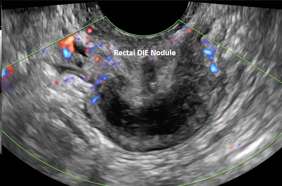
Endometriosis most commonly lies on the surface of pelvic tissues and organs, which is called Superficial Endometriosis. It can be readily seen and treated during laparoscopic surgery, but is poorly seen with Utrasound and MRI. Over time these superficial lesions can grow beyond the organ surfaces and invade the underlying tissue, which is then called Deep Infiltrating Endometriosis. When this happens, nodules will begin to form which can be challenging to identify during surgery. The nodules can show up on ultrasound if the examiner knows where and how to look for them. This requires a well trained sonographer who has extensive experience with the advanced techniques and imaging protocols that are required to accurately find and characterize the nodules in detail.
Deep Infiltrating Endometriosis most commonly affects the rectum, bowel, uterine ligaments, cervix, bladder, ureters, and vaginal wall. If left undetected it can lead to multiple and higher risk colorectal or urological surgeries. Identification of deep infiltrative lesions is critical for surgical planning because it helps define the complexity and associated risks of the procedure; better informing patients and improving their surgical outcome.

A typical pelvic ultrasound is effective for detecting ovarian endometriomas, which are blood filled cysts caused by endometriosis. However, a routine pelvic ultrasound will overlook sites of deep infiltrating endometriosis. MRI has been used to identify Deep Infiltrating Endometriosis but with limited results at 3 to 4 times the cost. A true Deep Infiltrating Endometriosis ultrasound scan protocol requires a detailed examination of structures that lie deep in the posterior pelvis, anterior and lateral compartments as well as the lower abdominal quadrants. Tis includes the rectum, cecum, terminal ileum and appendix, uterosacral and round ligaments, pouch of Douglas, vaginal wall, rectovaginal septum, ovarian fossa, ureters and bladder wall. An assessment of organ mobility is also performed to determine the presence and degree of adhesions.
